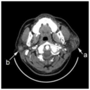
Mechanized tomography (CT) is obvious to the picture and shows the bone subtleties decisively dissimilar to attractive reverberation imaging, which portrays delicate tissues with high precision, and this method is liberated from torment.
What Is Computerized tomography
What Is Computerized tomography
What Is Computerized tomography
A CT filter is an X-beam restorative imaging gadget used to make a three-dimensional picture of the body's inner organs. It is framed by a few two-dimensional pictures taken around a fixed pivot of revolution.
Axial tomography
The data and pictures coming about because of a tomography gadget are handled to show the body's organs as per their capacity to forestall X-beams from going through them, and this data can likewise be utilized to draw three-dimensional pictures, and the pictures acquired are cross-sectional in nature, and every two-dimensional imaging slide is like what You see it when you cut an apple down the middle over the center and afterward you take a gander at the cutting surface.
On the off chance that you start that from the highest point of the apple down to the base, you will get a progression of cuts that speak to the all-out structure. With the assistance of a PC, 3D pictures of these two-dimensional slides can be reproduced.
Employments
Automated tomography is utilized to analyze different maladies, for example, harmful and considerate tumors in various territories of the body.
The mind
A CT filter is utilized to analyze tumors, inner dying, and calcifications. Tumors are effectively observable in light of the fact that they swell and increment in size, making harm the encompassing tissues. Ambulances are regularly outfitted with little CT scanners to check for stroke or a hit to the head.
An uncommon kind of these gadgets considered surgeries that are utilized when evacuating a tumor inside the head or treating arteriovenous abnormalities are frequently utilized. The cerebrum can likewise be filtered by an MRI gadget, yet a few infections and tumors must be seen through a tomography.
the chest
Electronic tomography is utilized to analyze and note changes in the inner light lung tissue. This sort of progress can't be seen with typical X-beams and MRIs. What's more, this review is utilized to discover harmful and different tumors in the lungs.
A CT check is regularly taken twice, once at inward breath and once upon exhalation. This procedure is called high-goals tomography.
It is utilized for blood vessel and venography imaging rather than interventional radiology and is more secure, brief timeframe and symptomatic of aspiratory embolism as opposed to atomic imaging.
PC tomography highlights
There are numerous qualities that improve this filtering technique than other medicinal examining strategies. To start with, tomography can show an away from the part being shot without indicating the organs that encompass it. For instance, while shooting the lungs, the heart or viscera doesn't show up in the image.
Second, the shading contrast between the tissues in the picture assists specialists with realizing the distinction in mass thickness. Also, this system can create great pictures without discharging a lot of radiation.
The most distinctive component of this system is that it isn't important to put any gas or gadget straightforwardly inside the body just like the case in the catheter.
To what extent does this assessment take
Present-day CT checks permit us to get pictures inside a couple of moments. On the off chance that intravenous infusion with a hued substance is required, this will require an extra time of close to a couple of moments too, and the assessment time frame, for the most part, goes between 15-20 minutes.
Degree of utilization
The utilization of registered tomography has expanded in the course of recent decades. In 1980, just 3 million registered tomography tasks were performed on patients, while in 2007 roughly 70 million figured tomography activities were acted in the United States of America. Nearly 6% of the imaging performed on youngsters.
This solid turnout isn't just in America yet in many nations of the world. The primary explanation is that most specialists want to play out a tomography of patients who are conceded direly to the clinic so as to precisely evaluate their wellbeing.
Additionally, one of the most significant reasons that make this photography so well known is its simplicity and speed. The photography procedure alone takes a couple of moments.
How is the photography procedure?
The gadget that issues the X-beam rotates around the segment to be imaged, and sensors or receptors get the radiation at the furthest edge of the circuit. These receptors contained xenon (Xe) however were later supplanted by increasingly compelling receptors.
Old imaging gadgets moved the article that was imaged a short while after the X-beam source machine had a total cycle, and the new gadgets permitted the body to move while the source gadget was turning around it.
Registered tomography is utilized in the restorative field as a demonstrative device, particularly before a medical procedure. Photographs taken must be handled before acquiring high-goals cross-sectional pictures of the part being shot.
The most effective method to print the picture
Pixel is a point of a specific shading in the 2-D picture. Voxel resembles a pixel, however in 3D. The shade of the pixel or voxel relies upon the porousness of the X-beam through the tissue being imaged. One transmittance is the Hounsfield unit.
The most porous tissues are given the estimation of 3070 units of Hounsfield, and the least penetrable ones are - 1024 units of Hounsfield. At the point when the pictures are printed, the tissues with pervasion of in excess of 80 units of Hounsfield show up in white, the tissues with saturation of under 0 units of Hounsfield show up in dark, and the tissues whose porousness is somewhere in the range of 0 and 80 units in Hounsfield show up in dim, the dimness of the dim shading identified with the level of penetrability. Likewise, when printing, the picture shows up topsy turvy.
The left piece of the picture speaks to the correct piece of the patient, and the other way around.
Arrangements before beginning the photography procedure
Before beginning the photography procedure, there are a few stages that should be taken. In the first place, the patient must educate the specialist in the event that he has any sensitivity to specific substances since he will be infused with a differentiation operator.
Second, the patient must educate the specialist in the event that he experiences past issues or infections identified with the heart, asthma, or others in light of the fact that these maladies may build the reactions of X-beams.
Third, the patient must quit eating a few hours before the assessment. It is desirable to wear agreeable and roomy apparel before processed tomography. At long last, the patient is required to take off adornments, gems, shoes, and pins, as this may unfavorably influence the assessment.
The most effective method to do this assessment
You will be approached to lie on a moving strong table. At that point, by moving the portable table, the piece of the body to be shot is put in a doughnut-like gadget. You will be approached to stay still as the scarcest development may influence picture quality. The imaging professional may request that you hold your breath to keep up the picture quality.
All through the sweep, the imaging professional screens you through a little window. There is additionally an interior specialized gadget that permits correspondence among you and the picture taker. Toward the finish of the assessment, you can return promptly to proceed with your standard work.
Now and again a pivotal tomography expert may choose to give you an intravenous infusion of shading material to explain what is found in the photos. At that point, the imaging specialist puts an intravenous needle through which to infuse the color. While infusing the color, you may feel a general warm inclination and a metallic preference for the mouth.
Now and again of imaging, the stomach related framework (belly), another sort of color might be mentioned by drinking so as to acquire an increasingly exact determination, as indicated by the case. This color is smashed one to four hours before the assessment, contingent upon its sort.
3D area pictures
The 2D pictures taken are regularly gathered and prepared by uncommon PC programming to get the 3D pictures. For instance, while shooting the spine, the two-dimensional picture shows just one passage of the spine, while the three-dimensional picture shows the situation of the vertebra according to the encompassing vertebrae and furthermore the ligament that ties the vertebrae to one another.
In the wake of handling the subsequent picture, the hues are added to it so as to encourage recognizing the various individuals. The point of these photos is to help specialists when performing medical procedures. The organs close to the spectator are normally semi-straightforward so they can see the other portion of the picture.
The negative impacts of PC tomography
X-beams utilized in tomography harm the cells in the body and atomic acids, which causes various tumors. Additionally, tomography utilized in diagnosing carcinogenic tumors may cause malignancy. The measure of radiation delivered by these gadgets is in excess of a thousand times the sum created by x-beams.
Likewise, numerous patients experience imaging more than would normally be appropriate, which builds their danger of creating malignant growth. Researchers recommend that roughly 5% of malignancy, later on, will be brought about by medicinal imaging of various types. The probability of creating malignant growth shifts as indicated by the individual's age.
The measure of radiation delivered while capturing youngsters can be diminished to secure them, as kids are bound to create malignancy contrasted with grown-ups. In spite of the fact that registered tomography has symptoms, it can't be shed or supplanted with other imaging strategies.
The measure of radiation and goals
There is an away from the connection between's the measure of x-beams gave by the gadget and the lucidity of the subsequent picture. The more the measure of beams, the more precise the picture is, and the other way around.
Nonetheless, given that these beams can cause malignant growth and hereditary imperfections, the measure of these beams must be held under a specific breaking point.
The measure of radiation can be diminished while keeping the picture goals worthy by following the accompanying strategies: 1 Changing the measure of radiation as per the age, weight, and sexual orientation of the patient and as indicated by the part to be captured.
2 Ensure that automated tomography is the most suitable strategy for the patient's condition. 3 Development of the product utilized in imaging to acquire more excellent pictures from lower x-beams.
Ages CT scanner
CT checks are ordered into a few ages as per the development of the filtering component, its speed and the time taken to frame the picture:
The original
The original CT scanners utilized a pencil-thick shaft coordinated to the body and checked by just a couple of identifiers.
The pictures are gathered through a rotational and transitional output where the X-beam source and the locator are introduced in a gadget called the gantry and pivot comparable to one another with the goal that the human body is in the hub of revolution for them, and the timeframe for one picture is assessed at 4 minutes where the gantry has made an entire 360-degree cycle Then, Gantry moves to check another piece of the human body, and the utilization of this age required submerging the patient's body in an aquarium to decrease its presentation to X-beams.
second era
The CT examine was grown so the number of reagents expanded and the X-beam bar turned out to be all the more wide to cover the relating reagents, the checking technique is as yet like the filtering strategy utilized in the original, and is by methods for a round and transitional sweep around the human body, an expansion in the quantity of reagents and an increment in the plentifulness of X-beams drove Until the filtering cycle for each area of the body secured 180 degrees with a change of 30 degrees rather than one degree as it was in the original, which brought about diminishing the filtering time.
third-era
There has been a perceptible improvement in the third era regarding speed in getting the picture, by dropping the transitional development and making the development round just, which made the examining time just one second.
To dispose of the transitional development during examining in the third era, the locators that screen the X-beams that are executed from the human body are planned as a circular segment, which keeps up a steady separation between the wellspring of the X-beams and the finders during the revolution.
Hindrances have additionally been included between the patient and X-beams, and between the patient and the reagents, to guarantee a dainty light emission beams that infiltrate the human body, in this way decreasing its presentation to radiation.
The fourth era
The fourth era was planned like the third era as far as filtering with a roundabout movement in particular, and the expansion that happened on the reagents that were introduced all through the whole circuit of the quantity of finders, which made the development confined to the X-beam source just with the security of the reagents since it encompasses the whole gantry.
This structure made checking a total segment of the body take close to one second, and right now gadget was imaged utilizing X-beams all through the district.
Modernized tomography or MRI
It is utilized for the essential assessment for bone or joint wounds for the most part X-beam assessment, it is quick and moderate. In cases that include head wounds, mechanized tomography is the primary decision, whereby it is conceivable to know the nearness of seeping in the mind or deferred skull inside minutes.
For the analysis of versatile tissue, MRI is utilized, for example, the recognition of ligament, ligaments, connective tissues, and muscle tissue. These tissues are portrayed by slight contrasts in thickness, which makes automated tomography doesn't give an exact analysis.
For finding in body regions, for example, the discovery of gallstones, changes in the liver or stones in the bladder, an ultrasound check is adequate.
Automated tomography depends on X-beam imaging, and MRI relies upon the utilization of the attractive field and electromagnetic radiation. And keeping in mind that the expense of a CT check is in the scope of a few a huge number of dollars, the expense of the MRI machine arrives at a large number of dollars.
Alert being used
One of the burdens of processed tomography is the moderately enormous portion of the radiation that the body is presented to. During it, the body is presented to a radiation sum proportional to multiple times the X-beam analysis on the chest. Identical to 50 mammograms.
In this way, the specialist must weigh between utilizing it due to its points of interest in exact conclusion and treatment, and the damages.
As indicated by the German Haier measurement: in 2003 electronic tomography was utilized in 6% of all findings by X-beams, however, it spoke to half of the restorative X-beams.
The United States of America directed around 52 million modernized tomographies, and experts gauge that 33% of those The conclusions were a bit much.
Radiation utilized in a CT output can crush the cells of the body, including DNA atoms, that can prompt malignancy.
References
Encyclopedia Wikipedia


