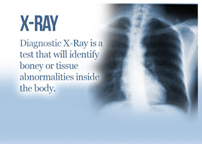
X-beams are an assessment during which the electromagnetic radiation gave by an extraordinary radiation gadget enters the body's tissues, hitting a plate set behind the body, to frame the picture of the body's organs that have been infiltrated by the beams.
What Is X-ray
What Is X-ray
X: ray are electromagnetic radiation with a wavelength (somewhere in the range of 10 and 0.01) nanometers, that is, the vitality of their beams is somewhere in the range of 120 and 120 thousand electron volts. It is broadly utilized in radiography and in numerous specialized and logical fields.
X-ray detection
William Roentgen, the X-beam pioneer, shed an electronic bar inside a glass tube applied between the two parts of the bargains pressure. This cylinder was vacuumed and electrons in it were discharged from the negative anode to the positive cathode. This cylinder is encased in light shading paper to shield the client from the transmitted electromagnetic field.
A luminous screen is put toward the finish of the cylinder. At the point when the electronic bar hit it, this screen began to gleam. When Richard Röntgen incidentally put his hand between the cylinder and the luminous screen, he saw an image of his hand bones on the screen, and this was the primary X-beam.
X-ray uses
Radiography in medication to distinguish teeth and bones and their breaks and find strong bodies, for example, shrapnel or lead in the body, just as identify tumors in the body, on account of these beams it is conceivable to consider unresolved issue with high exactness as these beams can enter the delicate bodies, for example, the skin however they can't Passing through the bone, which prompts the presence of the last picture. The most significant thing that recognizes it is the absence of symptoms.
Specialists likewise utilize these beams to treat and kill dangerous tumors. X-beams slaughter malignant growth cells and dispense with them, while solid body cells recapture essentialness after a brief period and come back to a sound recuperation.
X-beams were likewise utilized in the business to recognize the breaks and splits between the metal molds and wood utilized in the assembling of vessels, and the investigation of the retention range of these beams in the material additionally assisted with making X-beams an approach to distinguish the components engaged with the piece and examination of various materials.
Right now, beams are utilized to recognize every synthetic component. It has gotten conceivable to gauge the thickness of strong materials and sweep modern parts for absconds that are not observable to the unaided eye with these beams.
In the security field, X-beams are utilized to screen the travelers 'packs in air terminals for weapons or bombs.
In the study of examining strong items, as utilizing X-beam diffraction, it turned out to be evident that there is a particular balance in certain sorts of solids (precious stones), and that was the start of a monstrous beginning in considering the properties of solids and gem structure, and information on the nuclear structure of the components.
In the field of workmanship, it was utilized to recognize the techniques for painters and recognize genuine canvases and phony artistic creations, in light of the fact that the hues utilized in old compositions contain numerous metallic exacerbates that ingest x-beams, and the hues utilized in present-day artworks are natural aggravates that assimilate x-beams in a little rate.
X-ray reactions
There are two fundamental sorts of responses that we can acquire with X-beams that happen between electromagnetic waves and body tissue:
Photoelectric impact:
where the photon will be completely retained and the nuclear ionization brings about a total shutdown and doesn't leave the body and won't show up on the X-RayDetector.
Compton dispersing impact:
where the photon loses some portion of its vitality and alters its course.
X-ray severity
X-beams have a place with the ionizing radiation. That is, causing ionization of the medium in which it passes by isolating a few electrons in particles and atoms. It can cause changes in living cells that may prompt disease.
Along these lines, governments build up guidelines and laws identified with the utilization of X-beams, regardless of whether in medication or in the business and screen the recognition of those directions and rebuff the individuals who abuse the guidelines as per the laws built up right now.
Be that as it may, X-beams are likewise used to battle malignancy by method for concentrating X-beams on disease cells. DNA is a deoxyribonucleic corrosive in living beings that are exceptionally delicate to X-beams, as it turns out to be progressively harmed by expanding its assimilation of these beams.
That is, introduction to a little portion of those beams, regardless of how little, lies the probability of a living cell transforming into a dangerous cell, in light of the fact that the radiation will ionize the particles inside the body, which will make them flimsy
(precarious), and this procedure is actually the reason for the hazard to the patient as temperamental iotas will look for electrons in proteins and DNA to fill their circles and reestablish steadiness to themselves. This is the reason this probability of disease is considered when utilizing X-beams in the conclusion Or in treatment.
When all is said in done, pregnant ladies ought not to be presented to X-beams on the body, and alert ought to likewise be practiced against their utilization in youngsters, and they may cause sterility in people if the regenerative frameworks are presented to them.
Radiation dose
As it is the measure of radiation that the patient or radiologist is presented to, however, our essential objective is the patient in light of the fact that the specialist won't be true in the radiation room and won't be presented to the measure of radiation equivalent to the sum that the patient is presented to, as the more prominent the measure of radiation the more prominent ionization of the iotas and from them builds the probability of changes and tumors Precancerous.
The measure of radiation portion relies upon the associating medium, which is the human body, and in light of the fact that the human body is hilter kilter and comprises of numerous kinds of molecules that make up the tissues and interior organs; The radiation response for every district of the body will be unique. These distinctions are clarified by the accompanying:
Introduction
It is indicated by the image X, and it is estimated as far as the number of particles coming about because of introduction to EM radiation in a specific volume of air.
Its unit is Coulomb/kg, C/Kg which approaches 3876R. The measure of presentation diminishes with separation. That is, the more prominent the separation of the item from the source, the less presentation was by the reverse square law:
X (d2) = X (d1) * (d1/d2) ^ 2
Dose
It is signified by the image D, which is the measure of vitality that has been caught up in the body by the connections recently referenced. Its unit is rad.
1 and 2 are connected by one condition:
D = f * X, where f-factor is the transformation factor.
Proportionate portion
It is meant by image H, where the body is influenced by numerous kinds of radiation contrastingly when presented to a similar portion and this is the thing that depicts the proportional portion.
It is determined by the accompanying recipe:
H = D * Q
Its unit of rems is if the portion unit is rads, or Silevert (SV) if the portion unit is dim.
Where: Q (quality factor); And the X-beam is practically equivalent to one.
The successful portion
It is signified as Defective, where each uncovered piece of the body is given a specific weight.
Where Defective = aggregate (wi * Hi)
It ought to be aggregate (wi) = 1.
X-beam at the dental specialist
The dental specialist utilizes x-beams to film the teeth with a film and realize what is spoiled so as to make an arrangement for dental reclamation. Be that as it may, the advancement of the film takes some time. Notwithstanding the trouble of putting away and recording these movies.
So I built up the strategy for shooting with movies and started to be supplanted by advanced photography by computerized sensors. This strategy requires less introduction to the patient during the imaging procedure. The picture shows up straightforwardly on the screen.
In taking X-beams, care is taken to secure different pieces of the patient's body that are not inside the extent of assessment from lead radiation. Likewise, the medical attendant taking a shot at the x-beam machine leaves the room in the wake of setting up the patient for imaging and works the gadget from outside the room all together not to open herself to radiation.
Typically, every medical attendant behaviors functional preparing like clockwork for a time of one day, to reestablish her insight into the standards of radiation work and to present new things right now.
References
Encyclopedia Wikipedia


